
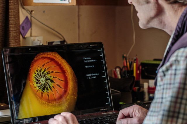
John W. showing me a 3D view of a peristome
After Jane, who came to stay with us last night, had left this morning to drive on to west Wales, Helena and I drove to the Museum in the Park to see an exhibition about the history of Stroud. We always like going to the Museum, which is sited in the old house complex at the centre of Stratford Park, and the exhibition was excellent. We both want to go back as there was so much to take in all at once.
Helena then went shopping for some last bits and pieces for her imminent trip to Scotland. I drove out of town up to Whiteshill to meet my old friend John W., as he had promised to show me some images he has photographed and processed for a recent demonstration. I have blipped John before and tried to describe some of the intricate work he does with microscopes and photography.
These particular images are made to show how quite commonplace objects from nature look when in extreme close up, and in 3D. John has found a way to photograph steroscopic images in his microscope and he can reproduce the stereoscopic images using software on his laptop and wearing radio controlled 3D glasses.
I took this picture of one of these images, this one being of part of a common type of water moss, called a 'peristome'. What you can see is the image which is 'presented' to one eye of a viewer. The image for the other eye is presented alternately at very high speed through the glasses, so that the composite image appears to be three dimensional. Filming the image requires extremely accurate photo-stacking of both the left and right hand images, which are then combined 'together' in software.
I saw about twenty of John's pictures, from a view of the 'sting' of a stinging nettle (horrifyingly vicious looking) to various forms and sizes of fungi, as well as things like lichens. One particular image showed how two different wing sections of a flying insect are shaped to act like velcro to enable the wings to be joined and then unjoined, with what appear to be lines of miniscule barbs. Seeing them so close up is quite astounding. This image shows a peristome which is approximately 1mm in total length, from the top of the laptop screen to the bottom, as indicated by the scale on the right of the screen. The hole at the top of it is where the spores of the fungus can be released from, the opening being adjusted or controlled by the degree of humidity, which is the controlling factor.
John can also answer questions about what it is you are seeing, because he is fascinated by natural systems and forms and is a wonderful teacher. I just wish that everyone could see these images for themselves. John loves showing them.
In case you are interested here is some information from Wiki about a Peristome.
Comments
Sign in or get an account to comment.


