
Inner Space
This bizarre-looking image was taken an hour ago using a very posh microscope.
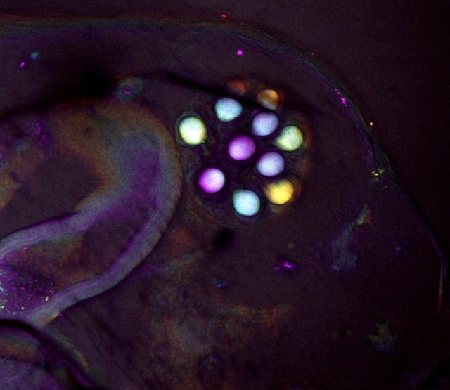
Two laser beams, one with a wavelength of 1064 nm and the other with a wavelength of 816 nm, were raster-scanned across a specimen - a small amount of the light that was back-scattered from the sample was blue-shifted relative to the incident beams. This blue-shifted light came most strongly from carbon-hydrogen molecular bonds, but also weakly from the water in the sample. So what you have here is a chemical map of anything in my sample that had carbon-hydrogen bonds or was wet.
The sample in question? It's an egg sac inside a Daphnia (a rather cute kind of crustacean).
If you can get your head around it, this image is only 530 microns across its longest side. 1 micron is one-millionth of a metre.
I've just got home after a very long day in the lab, but it was worth it. Simon has made me pasta and sauce for dinner :)
Edit to add (for the geeks): I applied a false-colour lookup table to a depth-stack taken through the sample, 5 slices with a step size of 5 microns, such that each slice had a different colour, then I produced a sum over all the slices to produce this multi-coloured image. The colours were: red, green, blue, indigo and violet - red is deepest into the sample.
423
views
- 1
- 0
Comments
Sign in or get an account to comment.


