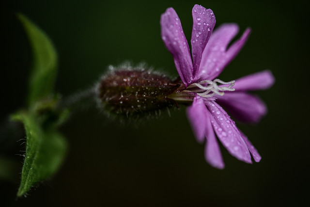
Campion
Closer in large ("L").
I had a brief photo-session in the early afternoon drizzle; my hectic week is now over (*phew*), although next week looks pretty mad too...
This working week ended with an hour-long demonstration of confocal microscopy which was absolutely amazing. It is a form of microscopy that uses a pinhole aperture to exclude light that isn't in focus, thus optically sectioning the specimen (imagine taking a photograph where only the in-focus section appears, and everything out of focus is excluded). We were shown a confocal laser-scanning microscope: we'd prepared specimens by adding fluorescent proteins (we injected transformed bacteria into plants, which then inserted genes that coded for fluorescent proteins causing transient expression of the fluorescent proteins) such that the laser could cause specific cell components to fluoresce, which were then visible in the microscope's output. The microscope doesn't work quite like a conventional one, but builds up images by scanning the laser across the specimen and recording the reflection: you can't look down it, but it produces images on a screen in (pretty much) real time.
We used stacks of optical sections (1 micrometre, 1 µm, thick: that is a millionth of a metre, or a thousandth of a mm) to generate three-dimensional views of individual cells (with the different components glowing in different colours), and also produced time-lapse videos of sub-cellular components whizzing around the cell.
It was wonderful; my macro images don't feel quite so close any more...
Comments
Sign in or get an account to comment.


